39 microscope labeled diagram
Neuron under Microscope with Labeled Diagram - AnatomyLearner Let's see the neuron histology slide labelled diagram and try to find out the below-mentioned characteristics - Presence of an identifiable cell body (soma) that locates in the brain's grey matter (according to the slide image). The cell body possesses spherical, euchromatic, and large eccentric nuclei containing a prominent nucleolus. Inverted Microscope- Definition, Principle, Parts, Labeled Diagram ... Light Microscope- Definition, Principle, Types, Parts, Labeled Diagram, Magnification Limitations Generally, these microscopes are very expensive to acquire. They are manufactured by very few companies because they are expensive to manufacture. They are rarely found in the market for purchase and usage.
Simple Squamous Epithelium under a Microscope with a Labeled Diagram ... Simple squamous epithelium under microscope labeled in renal corpuscle The cortex of a kidney consists of renal corpuscles and the convoluted tubule, straight tubules, nephrons, connecting tubules, and collecting ducts. You will find the medullary ray in the medulla of the kidney that comprises straight tubules and collecting ducts.

Microscope labeled diagram
Sperm Under Microscope with Labeled Diagram - AnatomyLearner Sperm Under Microscope 400X Labeled Diagram Before that, you may also read the below-mentioned article to get a full idea of the structure of seminiferous tubules - Histological features of the seminiferous tubules with the labeled diagram Okay, first, let's see the different histological features of the seminiferous tubules of an animal. Microscopy- History, Classification, Terms, Diagram - The Biology Notes History of Microscope. In the 1 st Century AD, the Romans invented the glass and used them to magnify objects. In the early 14 th Century AD, eyeglasses were made by Italian spectacle makers. In 1590, two Dutch spectacle makers, Hans, and Zacharias Jansen created the first microscope. It was a simple tube with 2 lenses system and had 9X ... Light Microscope- Definition, Principle, Types, Parts, Labeled Diagram ... Amazing 27 Things Under The Microscope With Diagrams Parts of a microscope with functions and labeled diagram 22 Types of Spectroscopy with Definition, Principle, Steps, Uses History of Microbiology and Contributors in Microbiology Microbiology of extreme environments (Types and Examples) Dark-Field Light Microscope
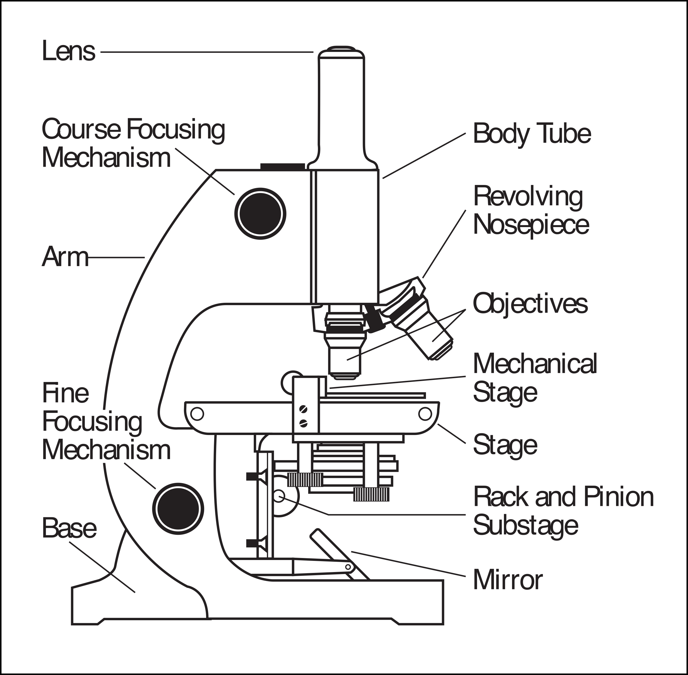
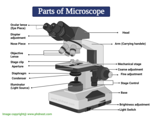
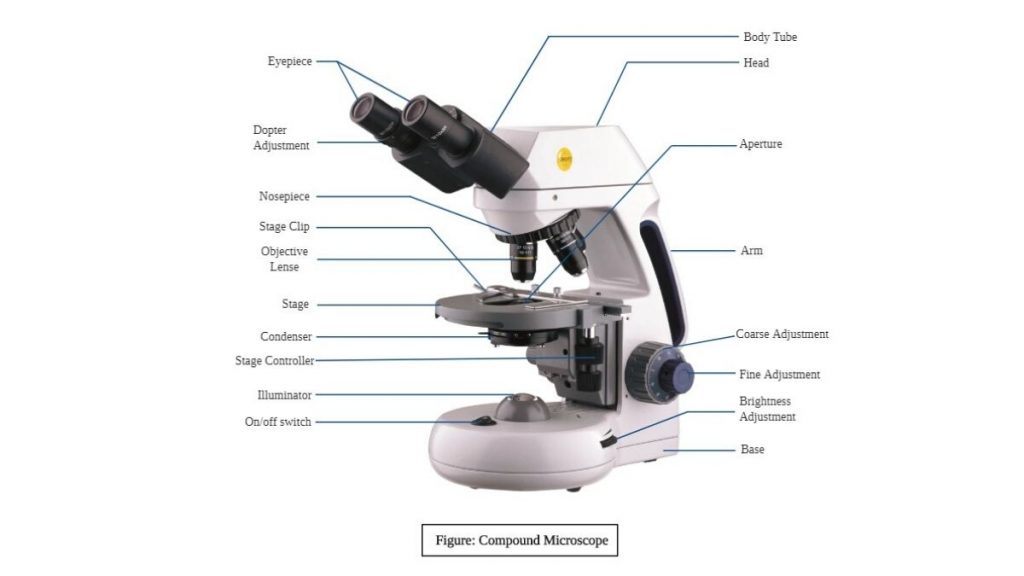
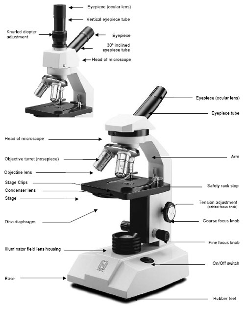
Microscope labeled diagram. Microscope: Types of Microscope, Parts, Uses, Diagram - Embibe There microscope anatomy includes three structural parts, i.e. head, base, and arm. Head - This is also known as the body; it carries the optical parts in the upper part of the microscope.. Base - It acts as microscopes support.It also carries microscopic illuminators. Arms - The microscope arm connects the base and the head and the eyepiece tube to the microscope base. Diatoms Under A Microscope Labeled - chunyinga.blogspot.com Microscope labeled diagram 1. They are generally of a golden-brown color and many are able to move about. What Are Diatoms Diatoms Of North America. Elegans under a stereo microscope. Full Hd Live Diatom Algae Under Microscope Magnification 400x. Pseudostratified Columnar Epithelium under a Microscope with a Labeled ... Pseudostratified Columnar Epithelium under a Microscope with a Labeled Diagram 06/04/2022 by anatomylearner The pseudostratified columnar epithelium comprises a single layer of cells but seems to be multilayered. It is because different cellular heights and nuclei are also placed at a different levels. Scanning Electron Microscope (SEM) - Diagram, Working Principle ... Scanning electron microscope is a classification of electron microscope that uses raster scanning to produce the images of a specimen by scanning using a focused electron beam on the surface of the specimen. An SEM creates magnified images of the specimen by probing along a rectangular area of the specimen with a focused electron beam.
Electron Microscope-Definition, Principle, Types, Uses, Labeled Diagram The electron microscope is placed vertically and has the shape of a tall vacuum column. It consists of the following elements: 1. Electron gun. A heated tungsten filament that produces electrons makes up the electron cannon. 2. Electromagnetic lenses. The condenser lens directs the electron beam to the specimen. Electron Microscope- Definition, Principle, Types, Uses, Labeled Diagram Electron Microscope- Definition, Principle, Types, Uses, Labeled Diagram April 4, 2022 by Sagar Aryal What is an Electron Microscope? Working Principle of Electron microscope Types of Electron microscope 1. Transmission Electron Microscope (TEM) 2. Scanning Electron Microscope (SEM) Parts of Electron Microscope 1. Electron gun 2. Compound Microscope- Definition, Labeled Diagram, Principle, Parts, Uses The naked eye can now view the specimen at magnification 400 times greater and so microscopic details are revealed. Alternatively, the magnification of the compound microscope is given by: m = D/ fo * L/fe where, D = Least distance of distinct vision (25 cm) L = Length of the microscope tube fo = Focal length of the objective lens Compound Microscope - Diagram (Parts labelled), Principle and Uses See: Labeled Diagram showing differences between compound and simple microscope parts Structural Components The three structural components include 1. Head This is the upper part of the microscope that houses the optical parts 2. Arm This part connects the head with the base and provides stability to the microscope.
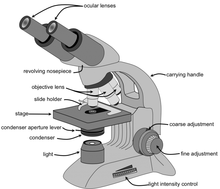
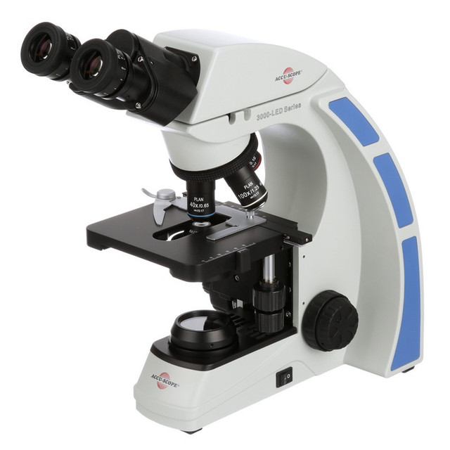
Testes: Anatomy, definition and diagram | Kenhub The testes (testicles) are male reproductive glands found in a saccular extension of the anterior abdominal wall called the scrotum. They are in ovoid shape, sized four to six centimeters in length. Testes develop retroperitoneally on the posterior abdominal wall and descend to scrotum before birth. The scrotum is often asymmetric, with one ... Microscope Types (with labeled diagrams) and Functions Simple microscope labeled diagram Simple microscope functions It is used in industrial applications like: Watchmakers to assemble watches Cloth industry to count the number of threads or fibers in a cloth Jewelers to examine the finer parts of jewelry Miniature artists to examine and build their work Also used to inspect finer details on products Microscope, Microscope Parts, Labeled Diagram, and Functions Microscope, Microscope Parts, Labeled Diagram, and Functions What is Microscope? A microscope is a laboratory instrument used to examine objects that are too small to be seen by the naked eye. It is derived from Ancient Greek words and composed of mikrós, "small" and skopeîn,"to look" or "see". Binocular Microscope Anatomy - Parts and Functions with a Labeled Diagram Now, I will discuss the details anatomy of the light compound microscope with the labeled diagram. Why it is called binocular: because it has two ocular lenses or an eyepiece on the head that attaches to the objective lens, this ocular lens magnifies the image produced by the objective lens. Binocular microscope parts and functions
22 Parts Of a Microscope With Their Function And Labeled ...
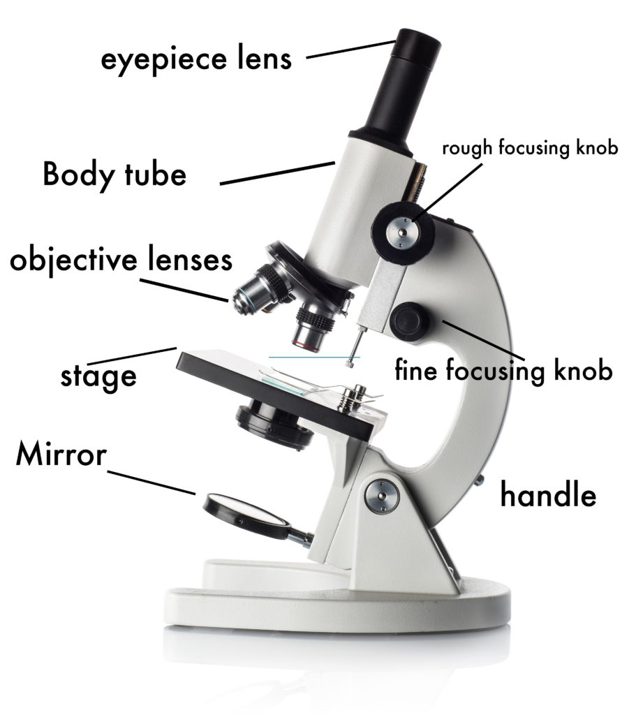
Parts of the Microscope with Labeling (also Free Printouts) Parts of the Microscope with Labeling (also Free Printouts) By Editorial Team March 7, 2022 A microscope is one of the invaluable tools in the laboratory setting. It is used to observe things that cannot be seen by the naked eye. Table of Contents 1. Eyepiece 2. Body tube/Head 3. Turret/Nose piece 4. Objective lenses 5. Knobs (fine and coarse) 6.

Microscope Terms Glossary | Earth science lessons, Medical ...
Simple Microscope - Parts, Functions, Diagram and Labelling Simple Microscope - Parts, Functions, Diagram and Labelling By Editorial Team March 7, 2022 A microscope is one of the commonly used equipment in a laboratory setting. A microscope is an optical instrument used to magnify an image of a tiny object; objects that are not visible to the human eyes. Table of Contents

microscope drawing with label - Clip Art Library
Light Microscope-Definition, Principle, Types, Parts, Labeled Diagram ... A light microscope is a device or instrument used in biology laboratories that uses visible light to locate, magnify, and expand micro objects. Using lenses, they focus light on the specimen and magnify it to generate a photograph. Typically, the specimen is positioned close to the microscopic lens. The kinds and quantity of lenses that make up ...

Microscope Parts and Functions
Bright-field microscope (Compound light microscope) - Diagram (Parts ... Bright-field microscope parts (Labeled Diagram) Ocular Lens This microscope has two eye lenses or ocular lens on the top of the microscope that are used to focus the image from the objective lens. It is from these lenses that we see the magnified image of the specimen. Objective Lens
Microscope parts labeled Icons PNG - Free PNG and Icons Downloads
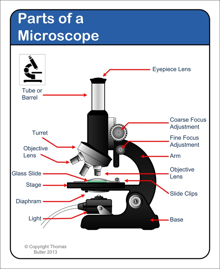
Microscope Parts, Function, & Labeled Diagram - slidingmotion Microscope parts labeled diagram gives us all the information about its parts and their position in the microscope. Microscope Parts Labeled Diagram The principle of the Microscope gives you an exact reason to use it. It works on the 3 principles. Magnification Resolving Power Numerical Aperture. Parts of Microscope Head Base Arm Eyepiece Lens

Parts of a microscope with functions and labeled diagram
Parts of a microscope with functions and labeled diagram - Microbe Notes Figure: Diagram of parts of a microscope There are three structural parts of the microscope i.e. head, base, and arm. Head - This is also known as the body. It carries the optical parts in the upper part of the microscope. Base - It acts as microscopes support. It also carries microscopic illuminators.

22 Parts Of a Microscope With Their Function And Labeled ...
Parts of a Microscope: Lesson for Kids - Study.com Now, look through the microscope like you would a pair of binoculars; these are the ocular lenses ('ocular' means eye). Sometimes when you look it's very dark. Slide your finger down to the base ...
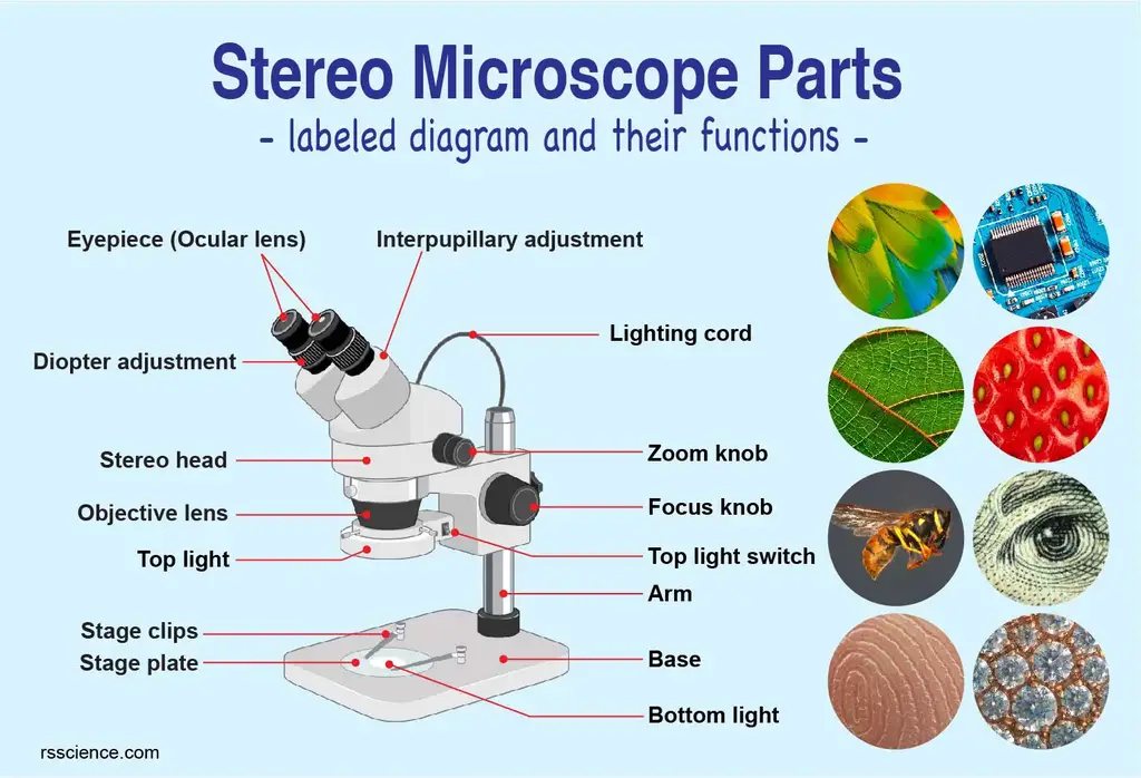
Parts of Stereo Microscope (Dissecting microscope) – labeled ...
Welcome to microscopy4kids - Microscopy4kids A microscope is an instrument to see objects that are too small to be seen by the naked eye. Its … Read More. Below is the most common resource about microscopes. Parts of Stereo Microscope (Dissecting microscope) - labeled diagram, functions, and how to use it. 10 Everyday Things You Should Look at Under a Microscope. Search. Search ...

Draw a well labelled diagram of a microscope. - Brainly.in
Simple Microscope - Diagram (Parts labelled), Principle, Formula and Uses Parts of a Simple Microscope A simple microscope consists of Optical parts Mechanical parts Labeled Diagram of simple microscope parts Optical parts The optical parts of a simple microscope include Lens Mirror Eyepiece Lens A simple microscope uses biconvex lens to magnify the image of a specimen under focus.

Labeled Microscope Diagram | Science fair projects ...
Microscope Diagram Worksheet - The Microscope Create A Labelled Diagram ... Microscope Labeled Diagram from cdn.slidesharecdn.com Used to support the microscope when carried. There is a printable worksheet available for download here so you can take the . This online quiz is called microscope labeling game science, microsope. Be sure to check our teachers notebook store for other printables.

29 | March | 2013 | IBG 102 Lab Reports
Microscope: Parts Of A Microscope With Functions And Labeled Diagram. Figure: A diagram of a microscope's components. The microscope has three basic components: the head, the base, and the arm. Head:Occasionally, the head is considered the body. It holds the optical components of the upper part of the microscope. Base:The microscope's base provides great support. It is also equipped with miniature illuminators.

Parts of Microscope, Function, Names & Labeled Diagram ...
Light Microscope- Definition, Principle, Types, Parts, Labeled Diagram ... Amazing 27 Things Under The Microscope With Diagrams Parts of a microscope with functions and labeled diagram 22 Types of Spectroscopy with Definition, Principle, Steps, Uses History of Microbiology and Contributors in Microbiology Microbiology of extreme environments (Types and Examples) Dark-Field Light Microscope
Welcome to Microbiology Lab King Saud University Dept
Microscopy- History, Classification, Terms, Diagram - The Biology Notes History of Microscope. In the 1 st Century AD, the Romans invented the glass and used them to magnify objects. In the early 14 th Century AD, eyeglasses were made by Italian spectacle makers. In 1590, two Dutch spectacle makers, Hans, and Zacharias Jansen created the first microscope. It was a simple tube with 2 lenses system and had 9X ...
Parts of Microscope, Microscope Labeled Diagram and Functions ...
Sperm Under Microscope with Labeled Diagram - AnatomyLearner Sperm Under Microscope 400X Labeled Diagram Before that, you may also read the below-mentioned article to get a full idea of the structure of seminiferous tubules - Histological features of the seminiferous tubules with the labeled diagram Okay, first, let's see the different histological features of the seminiferous tubules of an animal.
Microscope Types (with labeled diagrams) and Functions
How to Use a Microscope
Microscope - Teaching resources

Microscope Labelling Review Diagram | Quizlet
What are the main parts of a microscope? - Quora

Lesson Explainer: Microscopy | Nagwa
biology labeled microscope diagram - Clip Art Library

Compound Microscope Parts – Labeled Diagram and their ...
Compound Microscope Parts, Diagram Definition, Application ...

Parts of the Microscope with Labeling (also Free Printouts ...

Compound Microscope: Parts of Compound Microscope

Compound Microscope – Diagram (Parts labelled), Principle and ...
Compound Microscope Parts, Functions, and Labeled Diagram ...

File:Microscope diagram.png - Wikimedia Commons

Simple Microscope - Parts, Functions, Diagram and Labelling ...

Simple Microscope - Diagram (Parts labelled), Principle ...
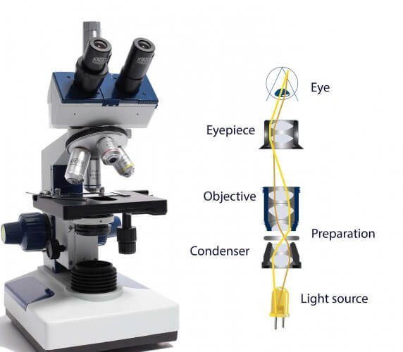
How do microscopes operate? - Krüss laboratory equipment

Label the microscope — Science Learning Hub
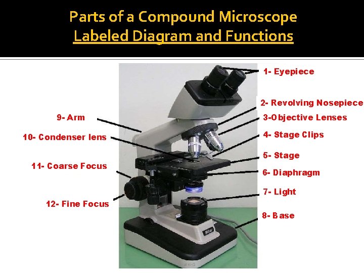
Parts of a Compound Microscope (And their Functions)

The Parts of a Microscope (Labeled) Printable Printable (6th ...
Microscope Maintenance Tips

2.1 " Compound Microscope" | Download Scientific Diagram

Microscope study part-2
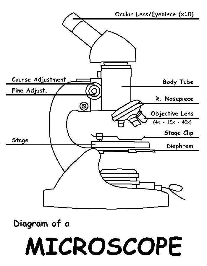
Diagram of a Microscope by ScienceDoodles on DeviantArt
1.2: Microscopes - Biology LibreTexts
Post a Comment for "39 microscope labeled diagram"